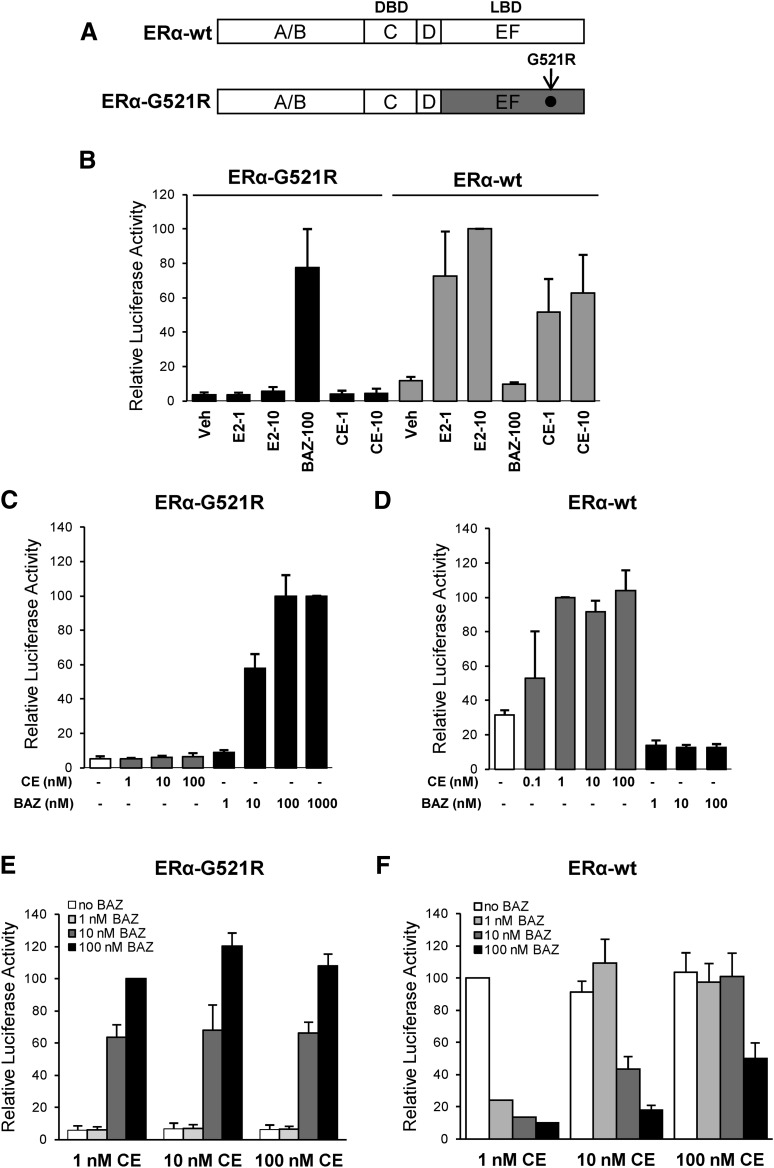
Fig. 1.
Activation of ERα-G521R with bazedoxifene. (A) Schematic diagram showing wild-type ERα (ERα-wt) and mutant ERα (ERα-G521R) used in these experiments. ERα consists of six domains (A–F). The A/B domains contain the ligand-independent AF-1 domain, the C domain is the DBD, D is the hinge, and the EF domain encompasses the LBD. The arrow indicates the position of the G521R point mutation. (B) HepG2 cells were cotransfected with 1 μg of pC3-Luc reporter and 50 ng of expression vectors for either ERα-G521R or ERα-wt. Cells were treated with the indicated concentrations of E2, BAZ, or CE for 24 hours, and then harvested for luciferase assay. The activity of 10 nM E2-treated ERα-wt transfected cells was set to 100. (C and E) HepG2 cells cotransfected with 1 μg of pC3-Luc reporter and 50 ng of ERα-G521R were treated with the indicated concentrations of ligands for 24 hours. The luciferase activity of cells treated with 1000 nM BAZ (C) or 100 nM BAZ + 1 nM CE (E) was set to 100. (D and F) HepG2 cells cotransfected with 1 μg of pC3-Luc reporter and 50 ng of ERα-wt were treated with the indicated concentrations of CE or BAZ alone or in combination for 24 hours. The luciferase activity of 1 nM CE treated cells was set to a value of 100 (D and F). Values represent the average ± S.E.M. of 3 independent experiments.

