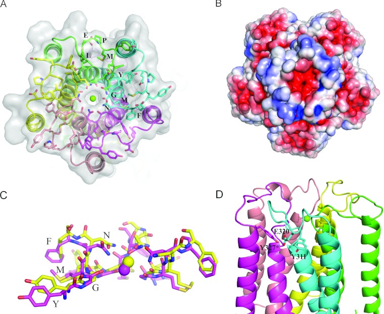
Figure 1. The spatial organization of the extracellular loop in the improved structure of TmCorA.
(A) The periplasmic loop of TmCorA is shown as sticks with the signature motifs YGMNF and MPEL indicated. Protein chains are colour-coded. The magnesium ion is drawn as a green sphere. (B) Electrostatic potential surface (±5 kT/e). Note the high negative charge at the selectivity filter. (C) Superimposition of YGMNF motifs from MjCorA (yellow) and TmCorA (magenta). RMSD (root mean square deviation) is ~0.6 Å. Three out of five monomers are shown. (D) The position of the charged Glu320 residue between two conserved tyrosine residues. *From adjacent monomer.

