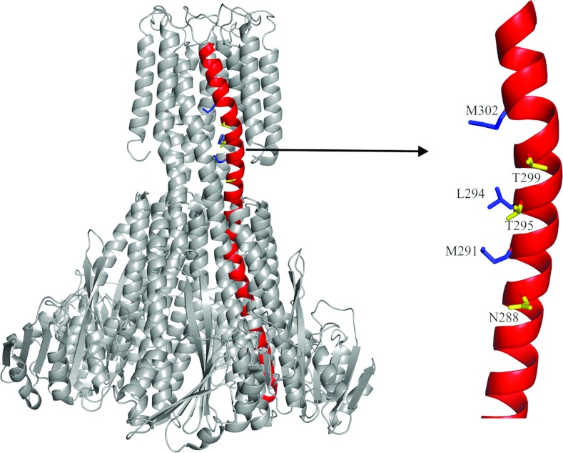
Figure 2. The arrangement of hydrophobic and polar residues on helix 7.
The structure of the pentameric TmCorA is presented in grey with helix 7 highlighted in red. The zooming of the transmembrane region of helix 7 shows the vertical alignment of both the hydrophobic residues (blue) exposed to the pore and the polar residues (yellow) facing away from the pore, in the closed conformation.

