
Figure 2.
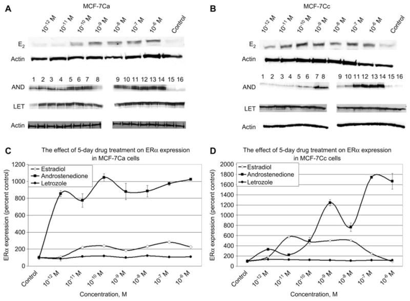
The effect of 5-day estradiol, androstenedione and letrozole treatment on ERα expression in MCF-7Ca and MCF-7Cc cells. (A,B) Western blotting analysis of ERα protein expression in nuclear extracts of MCF-7Ca and MCF-7Cc cells treated with either vehicle control (0.1% ethanol) or various doses of estradiol, androstenedione or letrozole for 5 days. Results of immunoblotting for estradiol treatment with anti-ERα is shown once whereas treatment with androstenedione and letrozole is shown in duplicate. (Lanes 1–2 represent 10−12 M; lanes 3–4 represent 10−11 M; lanes 5–6 represent 10−10 M; lanes 7–8 represent 10−9 M; lanes 9–10 represent 10−8 M; lanes 11–12 represent 10−7 M; lanes 13–14 represent 10−6 M; lanes 15–16 represent vehicle control) unless otherwise labeled. (C,D) Blots were striped and reprobed for β-actin and corrected for the internal loading control. Bands were quantitated by densitometry using Molecular Dynamics Software (ImageQuant) and values are represented as a percentage of the vehicle control±SEM (n=3).