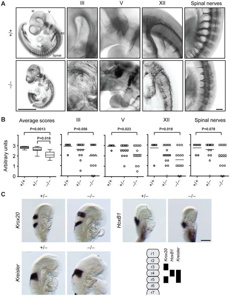
Figure 3. Cranial and spinal nerves are disorganized in Jarid1b knockout embryos.
(A) Embryos at E10.5 were stained with anti-neurofilament antibody. Shown are examples of a Jarid1b wild-type and knockout embryo (scale bar, 1 mm) and zoomed images (scale bar, 100 µm) of cranial nerves III, V and XII and spinal nerves. Note that the tail has been removed from the knockout embryo to facilitate imaging. (B) Scoring of nerve integrity: 3 = well defined; 2 = not very distinct, 1 = dysmorphic, 0 = absent. P values were determined by Mann-Whitney test. (C) Whole-mount in situ hybridization for Krox20, HoxB1 and Kreisler in E8.75 (5–12 somite pairs) Jarid1b heterozygous and knockout embryos. Scale bar, 200 µm. A scheme illustrating normal gene expression in the rhombomeric hindbrain is shown at the bottom right.

