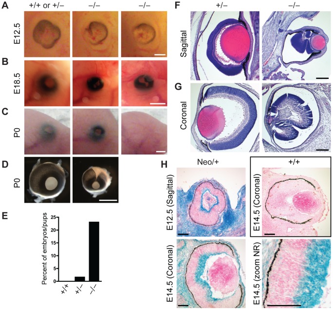
Figure 4. Defects in eye development in Jarid1b knockout embryos.
(A) External morphology of E12.5 eyes. Ventral is to the left. Note that the optic fissure is not completely closed in the Jarid1b knockouts while it is closed in controls (pooled +/+ and +/−). Scale bar, 200 µm. (B) Eyes of E18.5 embryos. Scale bar, 1 mm. (C) Eyes of newborn mice. Note that one of the knockouts has an open eyelid (left) while the other knockout completely lacks the eye (right). Scale bar, 1 mm. (D) Isolated eyes of newborn mice. Scale bar, 1 mm. (E) Frequency of the externally visible defects described in (B) and (C) for wild-types (n = 47), heterozygotes (n = 123) and knockouts (n = 78). (F, G) Hematoxylin and eosin staining of paraffin-embedded embryos at E18.5 showing the left eye cut sagittally (F) and the right eye cut coronally (G). Note that both knockout eyes are microphthalmic and the neural retina is misfolded. Scale bar, 100 µm. (H) Staining for β-galactosidase on sections of E12.5 and E14.5 eyes representing Jarid1b expression. For E14.5, a zoom on the neural retina (NR) is shown at the bottom right. Staining of a wild-type embryo is shown as negative control.

