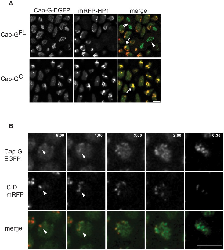
Figure 3. Cap-G-EGFP co-localizes with HP1 in interphase and initiates chromatin loading at centromeres.
(A) Cap-G is enriched at heterochromatic regions during interphase. Living embryos co-expressing UAST-Cap-GFL-EGFP or UAST-Cap-GC-EGFP (green in merged panels) and mRFP1-HP1 (red in merged panels) were analyzed while progressing through epidermal mitosis 14. Both EGFP-fused Cap-G-variants are locally enriched within interphase nuclei and show a particular co-localization with mRFP1-HP1 (arrows). Cap-GFL-EGFP localizes to the chromatin in metaphase (filled arrowhead) and anaphase cells (open arrowhead). (B) Cap-G loading initiates at centromeres. Embryos co-expressing gCap-GFL-EGFP (green in merged panels) and Cid-mRFP (red in merged panels) were analyzed to determine the initial sites of Cap-GFL-EGFP loading while progressing through post-blastodermal mitosis 14. Individual frames of a representative single nucleus are shown, with indicated times in min∶sec (t = 0, anaphase onset). Early Cap-GFL-EGFP accumulations frequently co-localize with Cid-mRFP1 signals (arrowheads). Scale bar 5 µm.

