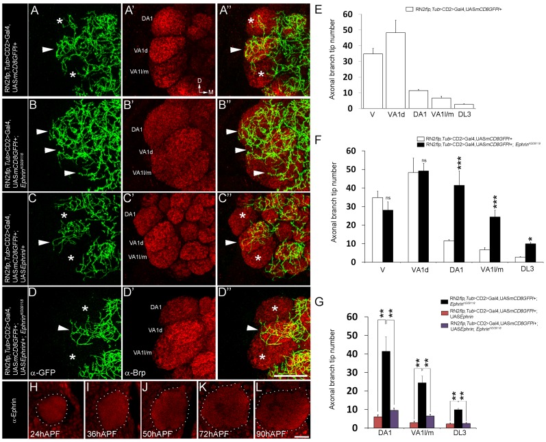
Figure 1. Glomerular-specific innervation pattern of the CSDn in the AL is regulated by Ephrin.
(A-A″, E) Innervation pattern of the axonal terminals of the CSDn (green) in glomeruli VA1l/m, VA1d and DA1 (anti-Brp in red) in control adults is shown (n>6). Asterisks indicate glomeruli with fewer innervations and arrowhead indicates glomerulus with more innervations from the CSDn. (B-B″, F) In EphrinKG09118 hypomorphs, increased terminal innervations can be seen to VA1l/m (n = 5, p<0.001), DA1 (n = 5, p<0.001) and DL3 (n = 9, p = 0.018) while innervations in VA1d (n = 5, p = 0.865) and V (n = 4, p = 0.149) are comparable to controls. (D-D″, G) Targeted expression of Ephrin in the CSDn in EphrinKG09118 hypomorphs restores distribution of axonal terminals in VA1l/m (n = 6, p = 0.99), glomerulus DA1 (n = 6, p = 0.606) and glomerulus DL3 (n = 6, p = 0.992). (C-C″, G) Targeted expression of Ephrin in the CSDn does not change overall distribution pattern of axonal tips in VA1l/m (n = 8, p = 0.241), DA1 (n = 8, p = 0.092233) and DL3 (n = 8, p = 0.910) when compared to controls, however a small decrease in overall branch tip number is observed. (E–G) Quantification of total axonal branch tip number in glomeruli V, VA1l/m, VA1d, DA1 and DL3 is plotted in histograms. A one-way repeated measure ANOVA test was performed to assess significant difference between the genotypes (F = 28.544, P<0.001). All pairwise multiple comparisions were performed using Fisher LSD method.. *, p<0.05; **, p<0.01; ***, p<0.0001; n.s. (not significant), p>0.05. (H–L) Ephrin shows broad expression pattern and it is expressed throughout the developing AL (n>5). APF = After puparium formation. All the images hereafter are oriented as indicated in A′ unless otherwise mentioned. D, dorsal; M, medial. Scale bar = 20 µm. See also Table S1.

