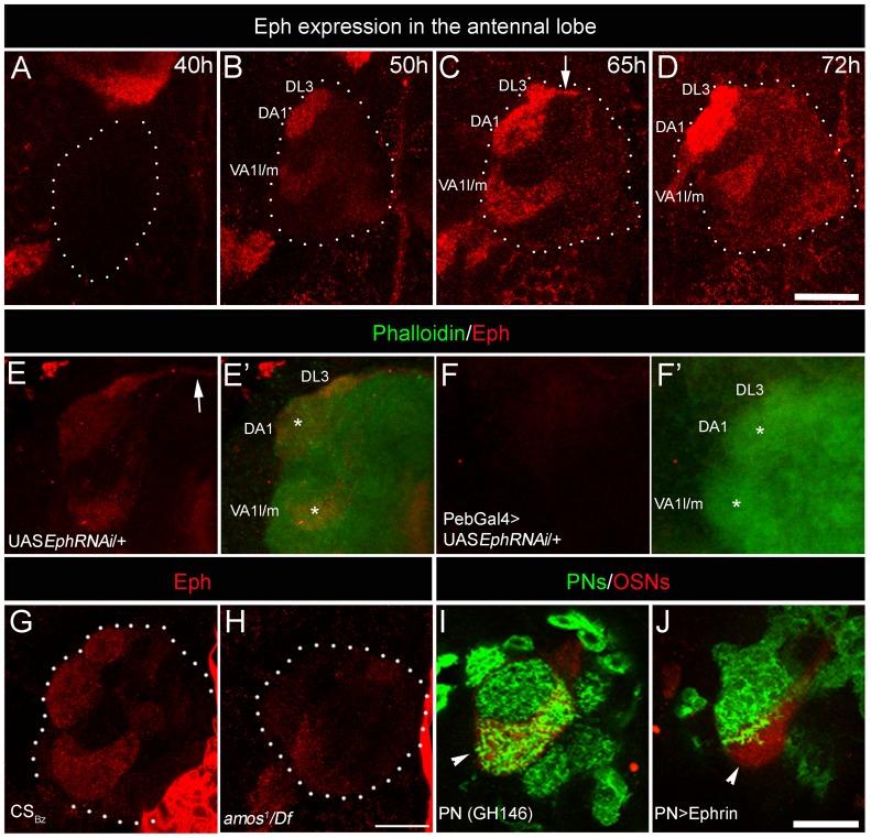
Figure 2. Sensory neurons differentially express Eph, which is capable of initiating repulsive interaction with Ephrin.
(A–D) Eph is strongly enriched in three anteriorly positioned glomeruli: VA1l/m, DA1 and DL3 glomeruli in the developing AL starting from 50 hAPF (n>5). White dots encircle the developing AL. Arrow indicates antennal commissure. (F-F′) Targeted expression of EphRNAi in sensory neurons (Pebbled-Gal4/+; UAS EphRNAi/+) results in strong reduction of Eph expression (red) in the antennal lobe compared to (E-E′) controls (UAS EphRNAi/+). AL is counterstained with phalloidin (green). (G–H) Eph expression is reduced in the AL of animals lacking majority of the OSNs from trichoid and basiconic sensilla. (G) Eph (red) is prominently expressed in select few glomeruli in the AL at 70 hAPF of control animals. (H) amos1/Df(2L)M36F-S6 animals show drastic reduction in the Eph expression in the AL. (I–J) Targeted expression of Ephrin in PNs prevents their entry in high Eph-expressing glomerulus VA1l/m (arrowhead). (I) In control animals (Gal4-GH146,mCD8::GFP/+; Or47b::rCD2/+), PN arbors (green) innervate glomerulus VA1l/m (red). (J) Very few PN arbors innervate VA1l/m glomerulus (red) when Ephrin is overexpressed in PNs (Gal4-GH146,mCD8::GFP/UAS Ephrin; Or47b::rCD2/+). Scale bar = 20 µm.

