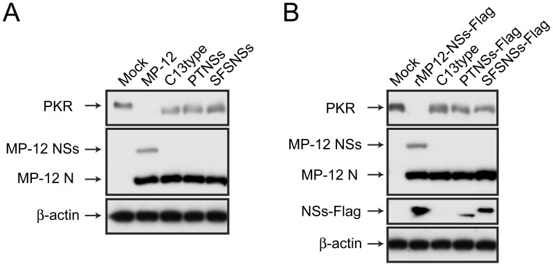
Figure 3. Degradation of PKR in VeroE6 cells infected with rMP12-PTNSs or rMP12-SFSNSs.
(A) VeroE6 cells were mock-infected or infected with MP-12, rMP12-C13type, rMP12-PTNSs or rMP12-SFSNSs at a m.o.i of 3, and cells were collected at 16 hpi. Western blot was performed with anti-PKR, anti-RVFV and anti-β-actin antibodies. (B) VeroE6 cells were mock-infected or infected with rMP12-NSs-Flag [32], rMP12-C13type, rMP12-PTNSs-Flag or rMP12-SFSNSs-Flag at a m.o.i of 3, and Western blot was performed as described above. Anti-Flag antibody was used for the detection of NSs-Flag. Representative data from three independent experiments are shown.

