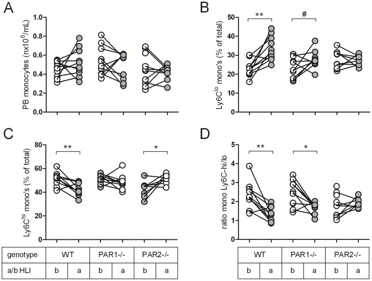
Figure 4. PAR2 contributes to the differentiation of circulating monocytes after femoral artery ligation.
Before (b, white circles) and seven days after ligation (a, grey circles), blood was drawn from WT, PAR1-/- and PAR2-/- mice to perform FACS analysis. (A) Absolute number of peripheral blood (PB) monocytes. Monocytes were gated to analyse the change in levels of (B) repair-associated Ly6C-low monocytes and (C) pro-inflammatory Ly6C-high monocytes upon ischemia, which is indicated in percentage of total monocytes. (D) Change in ratio of Ly6C-high/low monocytes upon ligation. # p<0.1, * p<0.05, ** p<0.01.

