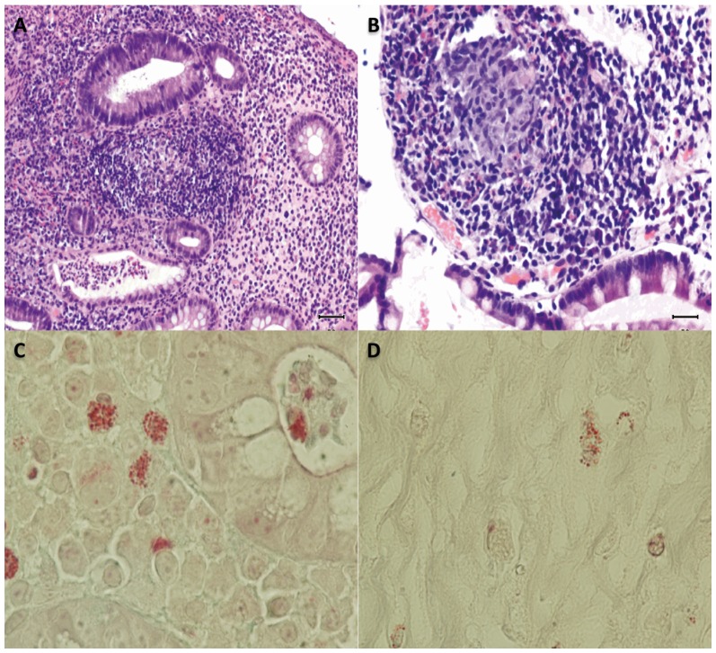
Figure 4. Tissue sampling of patient with CD.
A, Chronic inflammation with lymphoid nodules in the lamina propria with cryptitis and crypt microabscesses in some. Hematoxylin-eosin stain x 200. B, Structure composed of granulomatous histiocytic added. Hematoxylin-eosin stain x 200. C, Staining of undetermined structures in the macrophages next to a crypt gland. Modified Trichrome stain x 1000. D, Staining of undetermined structures in the macrophages next to a crypt gland in healthy control. Modified Trichrome stain x 1000.

