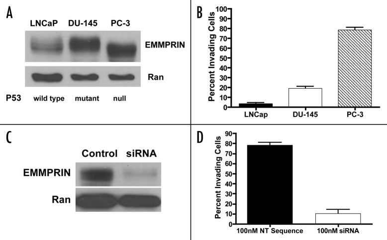
Figure 1. Expressions of EMMPRIN in human prostate cancer cell lines with different p53 status correlate with their respective invasive potential. (A) Cell lysates were prepared from LNCaP, DU-145 and PC-3 cells and equal amounts (50 μg) of proteins were resolved by 15% SDS-PAGE. Proteins were transferred to nitrocellulose membrane, and EMMPRIN was detected by immunoblotting with a monoclonal anti-EMMPRIN antibody. Ran was used as a loading control. (B) Cells suspended in media supplemente with 0.1% BSA were plated on Matrigel-covered filters in a Boyden chamber (3–5 x 10⁵ cells/well). Following incubation at 37°C for 48 h, the cells that reached the underside of the Matrigel-coated membrane were stained with hematoxylin and eosin and counted under a microscope at a magnification of x200. Each bar represents the mean ± SD of quadruplicate determinations from one of three independent experiments. (C) PC-3 cells in exponential phase of growth were transfected with an EMMPRIN-targeted siRNA (100 nM) or a non-targeting RNA. Forty-eight hours later, cell lysates were prepared from the transfected cells and EMMPRIN was detected as described above. (D) PC-3 cells in exponential phase of growth were transfected with an EMMPRIN-targeted siRNA (100 nM) or a non-targeting RNA. Forty-eight hours later, cells were plated on Matrigel-covered filters in a Boyden chamber (3–5 x 10⁵ cells/well). After incubation at 37°C for 48 h, invasive ability of the cells was determined as described above. Each bar represents the mean ± SD of quadruplicate determinations from one of three independent experiments.

An official website of the United States government
Here's how you know
Official websites use .gov
A
.gov website belongs to an official
government organization in the United States.
Secure .gov websites use HTTPS
A lock (
) or https:// means you've safely
connected to the .gov website. Share sensitive
information only on official, secure websites.
