
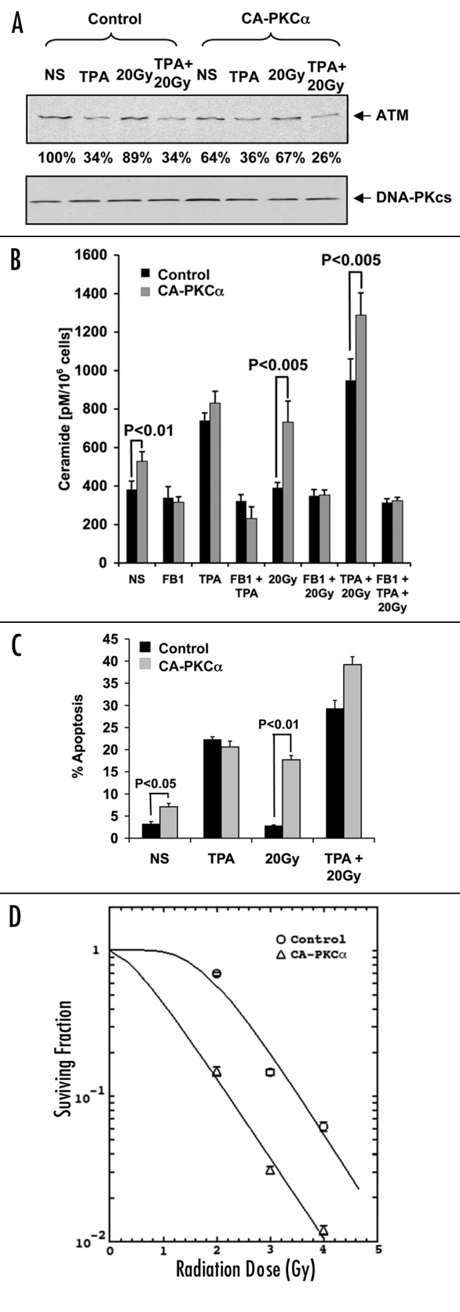
Figure 3. Effects of CA-PKCα on TPA-mediated apoptosis and ATM levels. LNCaP cells were transfected (t = 0) with 4 μg of CA-PKCα or control vector (and co-transfected with 0.4 μg GFP for apoptosis experiments), followed after 24 h with 20 Gy irradiation. (A) ATM expression was measured by western blot in transfected cells treated with 10 ng/ml TPA and/or 20 Gy of radiation. Sixteen hours following irradiation (t = 40 h), cell lysates were prepared. The results are representative of three experiments. (B) Measurement of ceramide levels in LNCaP cells (irradiated or non-irradiated) was performed 12 h post-irradiation (t = 36 h), as described in ‘Materials and Methods’. Values represent the mean average ± s.d. of triplicate measurements from two experiments. (C) Apoptosis of transfected cells treated with TPA and/or radiation (20 Gy). These experiments were performed as described in the legend of Figure 2. Results represent the mean average ± s.d. of triplicate measurements from three experiments. (D) LNCaP cells were transfected with either CA-PKCα or empty vector, incubated for 48 h and irradiated at various doses. The cells were subsequently replated at known concentration before culture for two weeks. Colonies of cells representing at least six divisions were scored. The curves were plotted using the single hit, multiple target (SHMT) model of radiation sensitivity. Each data point represents the mean average of six separate determinations, and show standard error.