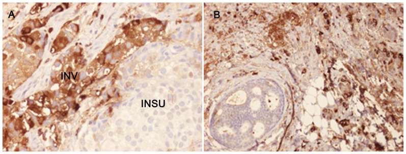
Figure 1. Immunohistochemical staining of breast tumour specimens showing the presence of IgA1 in the invasive part of the tumour.
A) In Sample 2, 40% of the tumour cells in the invasive part were stained with anti-IgA1 with a relative intensity of 3. Strong cytoplasmic and plasma membrane staining of IgA1 is observed in the invasive part of the section (INV) but only very weak staining in the in situ part (INSU). B) In Sample 7, 80% of the cells were IgA1-positive with a relative intensity of 3.

