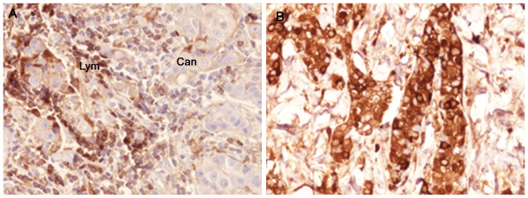
Figure 2. Immunohistochemical staining of specimens from individual breast cancer tumours showing the presence of IgA1 in both lymphocytes and tumour cells.
A) A section showing weak positive staining of cancer cells (Can) and intensively stained lymphocytes (lym). B) Intense anti- IgA1 staining of cancer cells in Sample 28, an ER/PGR-negative tumour in which 100% of the invasive part of the section was regarded as being maximally stained for IgA1 with an intensity of 3.

