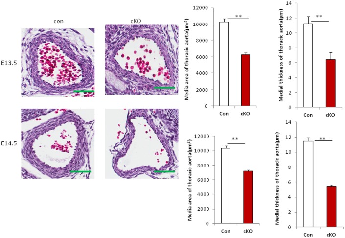
Figure 2. Loss of Drosha in VSMCs results in vascular wall hypoplasia in mice.
Sections of thoracic aorta from Drosha cKO and control embryos at E13.5 and E14.5 were stained with hematoxylin and eosin. The medial area and thickness of blood vessel walls of Drosha cKO mice and littermate controls were measured using the Elements image analysis program (Nikon) in three sections from each embryo. The media area of the vessel and the wall thickness were calculated from the inner and outer media circumference of vessel walls. Error bars indicate standard deviation (SD). Four different embryos were analyzed. Scale bars indicates 50 µm (**P<0.01; ***P<0.001).

