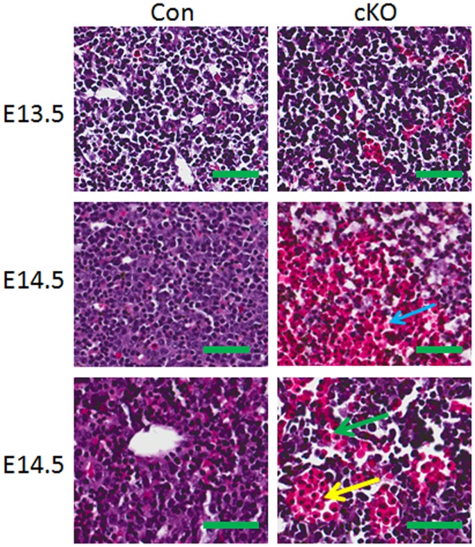
Figure 3. Drosha VSMC-specific cKO mutants display severe liver hemorrhage.
Sections from Drosha cKO and littermate control embryos at E13.5 and E14.5 were stained with H&E. The red blood cells occupied the hepatic plate (blue arrow), hepatic vein (yellow arrow), and sinusoids (green arrow) in the liver (scale bars represent 50 µm).

