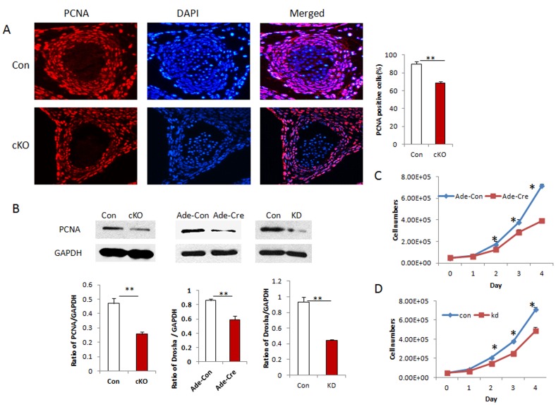
Figure 4. Disruption of Drosha in VSMCs of mice reduces cell proliferation.
A. Paraffin-embedded sections of thoracic aorta at E13.5 from control and cKO embryos were immunostained with the proliferating cell marker PCNA, and cell nuclei were counterstained with DAPI. The proliferating cells were counted and divided by the total number of nuclei as the proliferating index. Four different embryos were analyzed (error bar represent standard deviation; **P<0.01). B. VSMC proliferations in the umbilical arteries. KO VSMCs and KD VSMCs were examined using Western blot. The band intensity was normalized to GAPDH, and the ratios were used to analyze significant difference (**p<0.01, ***P<0.001). C. VSMC proliferation rates at different time points were examined by cell counts in Drosha KO and control VSMCs; rates were established by transducing Ade-Cre and Ade-Con viruses, respectively. Three separate experiments were performed, and significant differences were analyzed at each time point between control and KO cells (**P<0.01). D. Cell proliferations in KD and control VSMCs generated using retroviral knockdown vector were examined by counting cell numbers. Triplicate experiments were performed, and significant differences were analyzed between control and KD VSMCs (**P<0.01).

