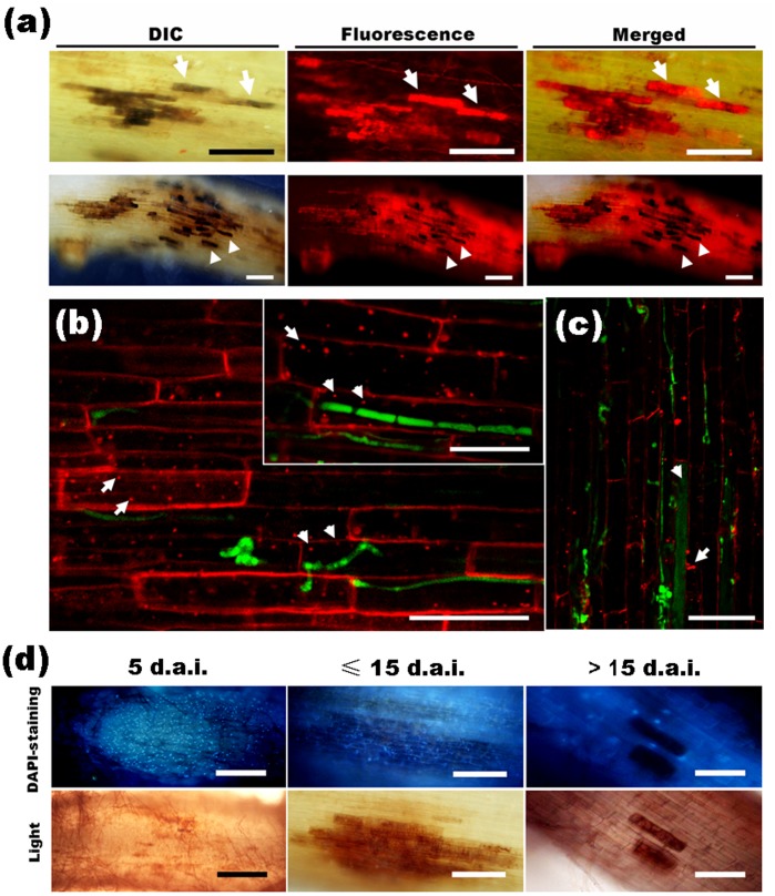
Figure 5. Vitality of H. oryzae-infected root cells as shown by staining with FM4-64 and DAPI, respectively.
(a) The colonized cells became melanized lesions and showed enhanced fluorescence (arrows) from the intracellular hyphae at 10 d.a.i. (upper panels). Bars, 50 µm. Later (≥ 15 d.a.i.), additional hyphae penetrated the root cells and the lesion-like infected area became larger; moreover, the enhanced fluorescence and plant autofluorescence of some primary infected cells disappeared (arrowheads) (lower panels). Bars, 200 µm. (b) The internalization of FM4-64 into endomembrane structures in fungus-infected cells (arrowheads) and non-invaded cells (arrows) during early colonization (≤ 10 d.a.i.). Bars, 100 µm. (c) At 15 d.a.i., endocytosis disappeared in the cells occupied by microsclerotia (arrowhead), but remained in the adjacent non-infected cells (arrow). Bar, 50 µm. (d) Root segments stained with DAPI. A large number of DAPI-positive nuclei in root cells with slight infection at 5 d.a.i. (left panels; Bars, 400 µm). DAPI-stained nuclei in root cells occupied by abundant hyphae and microsclerotia at 15 d.a.i. (middle panels; Bars, 200 µm). DAPI-stained nuclei disappeared in root cells filled with microsclerotia, but remained in the adjacent non-invaded cells after 15 d.a.i. (right panels; Bars, 100 µm).

