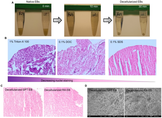
Figure 3. Decellularization of SPT and RA treated embryoid bodies.
(A) Panel images depict the decellularization process of EB. (B) Histological analysis of decellularized EB by H&E staining to show cell removal efficiency with three different detergents. (C) H&E staining of both groups of decellularized EB scaffolds showing absence of intact nuclei. (D) SEM images after decellularization process shows dense particulate material without distinct individual cell in both groups of EBs.

