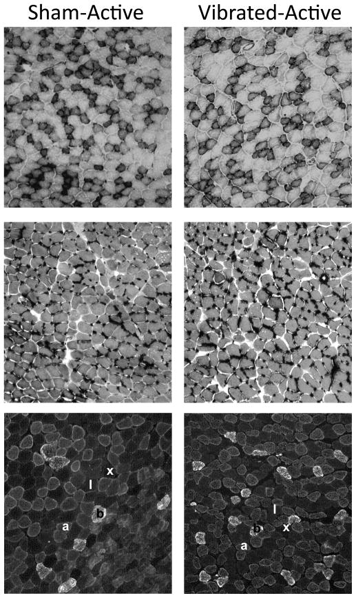
Figure 4.
Representative cross-sections of soleus muscles from mice that were caged in normal sized cages (Active) and were subjected to low intensity vibration training (Vibrated) or not (Sham). Top panel shows muscles stained for nicotinamide adenine dinucleotide (NADH)-tetrazolium reductase reactivity as an indicator of mitochondrial enzyme activity. Dark fibers were counted at positive. Middle panel shows muscles stained by periodic acid-Schiff reaction that labels capillaries. Bottom panel shows muscles triple-stained with antibodies against type 1, 2a, and 2b myosin heavy chain. Fibers denoted with “1” were classified as type 1 fibers, “a” as type 2a, and “b” as type 2b. These fibers were distinguished based on secondary antibodies that fluoresced fibers red, green, or blue. Fibers denoted by “x” were classified as type 2x because they did not react with any myosin heavy chain antibody and thus did not fluoresce.

