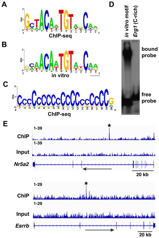
Figure 6. DMRT1 DNA association in the fetal gonad.
(A) Enriched motif in top 100 peaks, closely resembling in vitro defined DMRT1 recognition sequence. (B) In vitro-defined DNA binding motif. (C) C-rich motif enriched in DMRT1 binding peaks. (D) Gel mobility shift assay showing that DMRT1 can bind to the in vitro defined DMRT1 DNA binding motif but not to a C-rich sequence (5′TCCTCCCCCTCCTACCCCCCCCCCCCACAC3′) derived from a prominent DMRT1 binding peak in the Erg1 gene. (E) ChIP-seq data showing binding of DMRT1 to Nr5a2/Lrh1 and to Esrrb in the E13.5 testis. Binding peaks called by MACS are indicated by asterisks. Each peak contains a close match to the DMRT1 consensus DNA binding motif.

