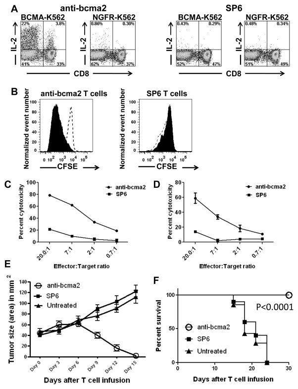
Figure 4. T cells expressing anti-BCMA CARs produced cytokines, proliferated, and killed in a BCMA-specific manner. Anti-BCMA CAR-transduced T cells eradicated tumors in vivo.
A, T cells from Myeloma Patient 4 were transduced with either anti-bcma2 or the negative-control CAR SP6. Four days later, the CAR-transduced T cells were cultured for 6 hours with either the BCMA-expressing cell line BCMA-K562 or the BCMA-negative cell line NGFR-K562. Large fractions of the T cells transduced with anti-bcma2 produced IL-2 when cultured with BCMA-K562. Only small numbers of anti-bcma2-transduced T cells produced IL-2 when they were cultured with NGFR-K562. Only small numbers of SP6-transduced T cells produced IL-2 when cultured with either BCMA-K562 or NGFR-K562. The plots are gated on CD3+ lymphocytes. The numbers on the plots are the percentages of cells in each quadrant. This is one of three experiments with similar results. B, T cells from Donor B were transduced with either anti-bcma2 or SP6. The T cells were then labeled with CFSE. The T cells were cultured with either irradiated BCMA-K562 cells or irradiated NGFR-K562 cells. IL-2 was not included in the cultures. Four days later, the T cells were analyzed by flow cytometry. The plots are gated on CAR-expressing T cells. The CFSE fluorescence was less intense in anti-bcma2-expressing T cells that were cultured with BCMA-K562 cells (solid) than in anti-bcma2-expressing T cells that were cultured with NGFR-K562 cells (dashed, open). This indicates that anti-bcma2-expressing T cells proliferated specifically in response to BCMA. For SP6-expressing T cells, CFSE fluorescence intensity was similar for T cells cultured with either BCMA-K562 or NGFR-K562. This is one of two experiments with similar results. Anti-bcma2-transduced T cells from Donor A specifically killed the multiple myeloma cell lines C, H929 and D, RPMI8226 in 4-hour cytotoxicity assays at various effector:target cell ratios. T cells transduced with the negative control CAR SP6 caused much lower levels of cytotoxicity at all effector:target ratios. For all effector:target ratios, the cytotoxicity was determined in duplicate, and the results are displayed as the mean +/− the standard error of the mean. E, Mice were injected intradermally with RPMI8226 cells and tumors were allowed to grow for 17 to 19 days. On day 0, the mice received intravenous infusions of 8×10⁶ T cells that were transduced with either anti-bcma2 or the negative control CAR SP6. Another group of mice was left untreated. The mice all had established tumors at the time of T-cell infusion. One-hundred percent of mice receiving infusions of anti-bcma2 had rapid and complete regressions of their tumors. In contrast, all mice receiving infusions of SP6-transduced T cells and all mice left untreated had progressive enlargement of their tumors. F, Tumors did not recur in mice receiving anti-bcma2-transduced T cells for the duration of the experiment. All mice receiving anti-bcma2-transduced T cells survived and were healthy for the duration of the experiment. All mice receiving SP6-transduced T cells or left untreated died with progressive tumors. The P<0.0001 refers to the comparison of anti-bcma2 and SP6. E and F show combined results of 3 experiments (anti-bcma2, n=10; SP6, n=10; untreated, n=7).

