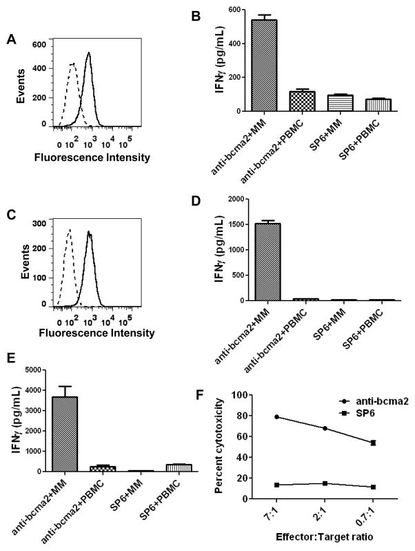
Figure 5. Anti-BCMA-CAR-transduced T cells specifically recognized and killed BCMA-expressing primary multiple myeloma cells.
A, Flow cytometry staining for BCMA (solid line) and isotype-matched control staining (dashed line) revealed BCMA expression on the surface of primary bone marrow multiple myeloma cells from Myeloma Patient 5. The plot is gated on CD38high CD56+ plasma cells, which made up 40% of the bone marrow cells. B, Unmanipulated myeloma-containing bone-marrow cells (MM) from Myeloma Patient 5 or PBMC from Myeloma Patient 5 were cocultured overnight with allogeneic T cells from Donor C. The T cells had been transduced with either anti-bcma2 or the negative control CAR SP6. After the coculture, an IFNγ ELISA was performed. C, Flow cytometry for BCMA (solid line) and isotype-matched control staining (dashed line) revealed BCMA expression on the surface of MM cells from a plasmacytoma of Myeloma Patient 1. The plot is gated on plasma cells, which made up 93% of the total plasmacytoma cells. D, Unmanipulated myeloma cells (MM) from a plasmacytoma of Myeloma Patient 1 or PBMC from Myeloma Patient 1 were cocultured overnight with allogeneic T cells from Myeloma Patient 4. The T cells were transduced with either anti-bcma2 or the negative control CAR SP6. The PBMC from Myeloma Patient 1 did not contain BCMA+ cells. After the coculture, an IFNγ ELISA was performed. E, Unmanipulated myeloma cells (MM) from a plasmacytoma of Myeloma Patient 1 or PBMC from Myeloma Patient 1 were cultured overnight with autologous anti-bcma2-transduced T cells or autologous SP6-transduced T cells, and an IFNγ ELISA was performed. F, Myeloma cells from a plasmacytoma of Myeloma Patient 1 were specifically killed by autologous anti-bcma2-transduced T cells at low effector:target ratios while autologous SP6-transduced T cells caused only a low level of cytotoxicity of the myeloma cells in a 4-hour cytotoxicity assay. For all effector:target ratios, the cytotoxicity was determined in duplicate, and the results are displayed as the mean +/− the standard error of the mean.

