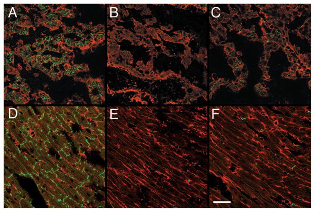
Figure 2.
Immunofluorescent staining for Cx43 in wild-type and MHC-CKO hearts. Sections from E12.5 (A through C) and 4-week-old (D through F) hearts were stained for Cx43 (green fluorescent stain), counter-stained using wheat germ agglutinin (red fluorescent stain), and imaged with confocal microscopy. Abundant Cx43 staining in control littermate hearts is evident at both embryonic (A) and postnatal (D) stages. In contrast, extensive areas of the myocardium in embryonic (B and C) and post-natal (E and F) CKO mice are devoid of Cx43 staining, although some residual expression is detectable. Bar=50 μm.

