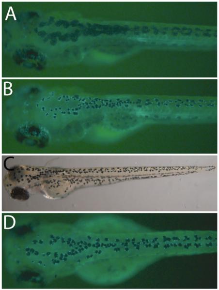
Fig. 2.
Techniques for visualizing individual melanocytes. (a) Wild-type zebrafish at 5 dpf expressing fTyrp1 > eGFP transgene. (b) 10 min epinephrine treatment (5 mg/mL) of fish shown in (a), which facilitates counting cells and clearly shows cells expressing GFP. (c) The mlphaj120 mutant line has constitutively contracted melanocytes that facilitate counting cells. (d) The fTyrp1 > eGFP transgene on a mlphaj120 background allows unambiguous identification of GFP + melanocytes.

