
Fig. 3.
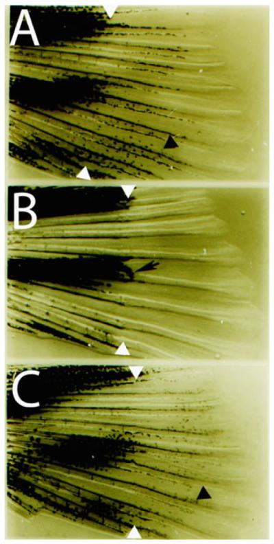
Reverse labeling of melanocytes using PTU. (a) Fin regeneration in an untreated caudal fin. White arrowheads indicate amputation plane and black arrowhead indicates regenerated pigmented melanocyte. (b) Fin regeneration in a PTU treated caudal fin. Note the lack of pigmented melanocytes in regenerate, except for a few melanocytes immediately distal to the amputation plane that preceded the amputation (black arrow). (c) Washout of PTU following fin regeneration (similar to b) reveals the presence of melanocytes (black arrowhead) that develop from unpigmented precursors.