
Fig. 4.
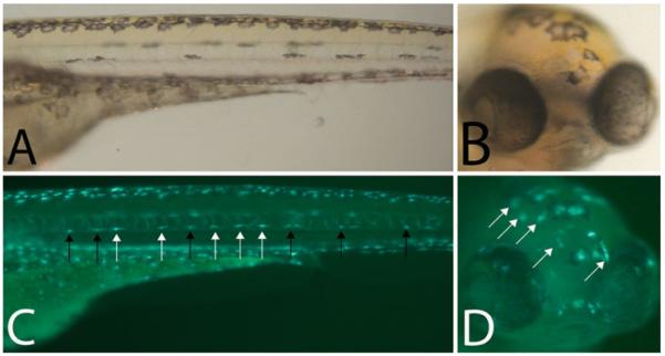
Birthdating melanocytes using Tg(fTyrp1 > eGFP)j900 in conjunction with PTU (a) Trunk and (b) head of 5 dpf zebrafish larvae treated with PTU at 2.5 dpf, blocking melanin synthesis in MSC derived embryonic melanocytes in the lateral stripe. (c, d) Epifluorescence of (a, b) reveals additional melanocytes that are not visible in bright field. Black arrows are previously differentiated melanized, GFP + melanocytes. White arrows are more recently differentiated unmelanized, GFP + melanocytes.