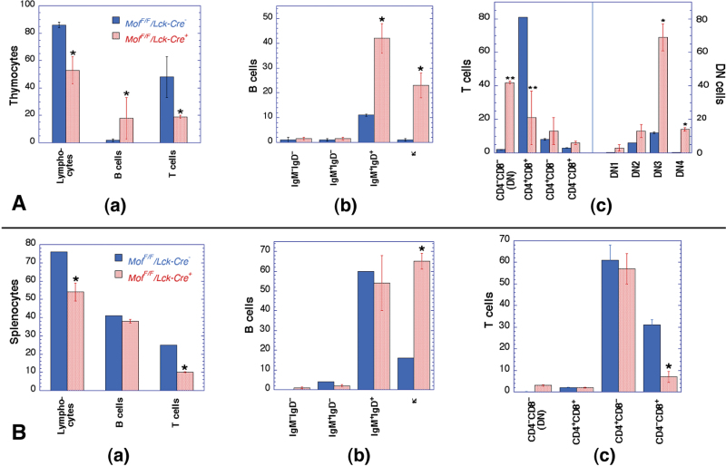
Fig. 3.
Analysis of thymus and spleen lymphocytes in Mof F/F /Lck-Cre + and Mof F/F /Lck-Cre – mice. (A) Comparison of lymphocytes from (a) Mof F/F /Lck-Cre + thymus indicating a significant reduction in total lymphocytes and pan T cells (CD3+CD5+) but increased pan B cells (B220+) levels; (b) thymic B cells from Mof F/F /Lck-Cre + mice indicate a significant increase in mature B cells (IgM+IgD+) with an unusually high clonal (κ light-chain) expansion ratio (κ); and (c) thymic T cells from Mof F/F /Lck-Cre + indicating an early developmental defect, increased population of CD4–CD8– (DN) T cells as well as statistically significant decreased CD4+CD8+ T cells. DN population was further classified into DN1 (CD44+CD25–), DN2 (CD44+CD25+), DN3 (CD44–CD25+) and DN4 (CD44–CD25–) populations and T cells from Mof F/F /Lck-Cre + accumulated in DN3 and DN4. (B) Comparison of cells from spleen (a) statistically significant reduction in total lymphocytes and T cells, whereas total B-cell number was unchanged; (b) difference in B cells, mature B cell (IgM+IgD+) was unchanged; however, abnormal κ light-chain clonal expansion was observed; (c) difference in splenic T cells, the number of helper T cells (CD4+CD8–) was unchanged; however, cytotoxic T-cells (CD4–CD8+) level was reduced. *P < 0.05 and **P < 0.001 determined by the chi-square test.

