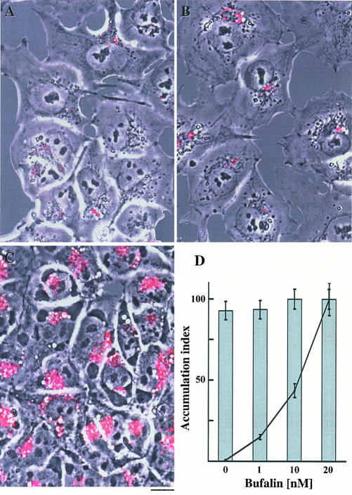
Figure 2.
Bufalin-induced translocation of cell surface fluorescence labeling into large perinuclear vesicles in NT2 cells. NT2 cells were grown in DMEM-F12 on glass coverslips for 24 h. The DMEM-F12 was then replaced with drug-free medium (A), or medium containing 15 μM etoposide (B) or 20 nM bufalin (C). After 20-min incubation, the medium was removed and the cells were incubated for 5 min in PBS containing FM1-43. The reagent was then removed; the cells were washed twice with PBS and incubated for an additional 4 h in the presence of the drugs mentioned above. All these procedures were carried out at 37°C in 5% CO2. Images were obtained and analyzed, as described in MATERIALS AND METHODS, and the fluorescence intensity/cell (solid line) and the percentage of fluorescent cells (bars) was calculated. The data obtained from 800 to 1200 cells were collected, and the average value ± SD was plotted (D). Bar, 10 μm.

