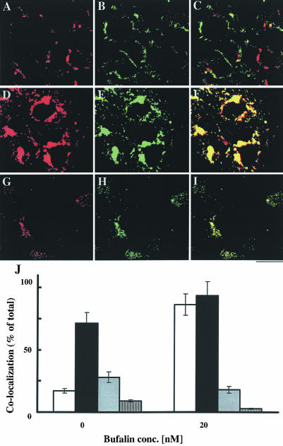
Figure 7.
Most of the transferrin that accumulates in bufalin-treated cells is associated with Rab11 and Rab7 proteins. Rab proteins and transferrin were detected as described in MATERIALS AND METHODS. The effect of bufalin on the colocalization of transferrin and Rab signals, and their quantification, were determined as described in Figure 5. Results obtained from the control cells (A-C) and from the bufalin-treated cells (D-I) are shown. The red color corresponds to the transferrin signals (A, D, and G), the green color to Rab11 signals (B, E, and H), and the yellow color corresponds to the merged images (C, F, and I). Images A-F were obtained by conventional fluorescence microscopy, whereas images G-I were acquired by confocal microscopy (0.4-μm sections). Bar, 20 μm. Quantification of the colocalization of transferrin with Rab4 (gray bars), Rab5 (dashed bars), Rab7 (solid bars), and Rab11 (white bars) is depicted in J.

