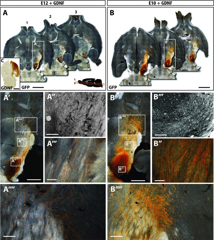
Figure 2. GDNF over-expression promotes the integration of both older and younger age donor grafts.

A and B, dark-field photomontages providing a horizontal overview of the GFP+ grafts of E12 (A) and E10 (B) donor grafts in the presence of GDNF over-expression, at 12 weeks post transplantation. C, GDNF was over-expressed by injection of AAV-GDNF into the host striatum at the time of grafting. Immunohistochemistry revealed robust GDNF expression throughout the target striatum and overlying cortex. Furthermore, GDNF was expressed within the efferent targets of striatal and cortical projection neurons, including the globus pallidus and substantia nigra. A′ and B′, magnified overviews of the E12+GDNF and E10+GDNF grafts localized within the VM, their trajectory through the medial forebrain bundle and targeting the striatum. Bright field images illustrating larger grafts within notably more GFP+ cells and increased cellular density in animals grafted with E10 donor tissue (+GDNF, A′), compared to E12 (+GDNF, B′) donor grafts. However, grafts were not notably larger than E10 or E12 grafts in the absence of GDNF (Fig. 1). A″′ and B″′, exposure of grafts to GDNF resulted in a marked increase in GFP+ fibres coursing through the MFB in both young and older donor grafts (compared to the absence of GDNF, Fig. 1). A″″ and B″″, additionally GDNF increased striatal innervation. The effects of GDNF on axonal growth and striatal targeting were more evident in younger than older donor grafts. AC, anterior commisure; CPu, caudate putamen; GP, globus pallidus; MFB, medial forebrain bundle; T, intranigral transplant. Scale bar: A,B= 2μm; A′, A″, A″″, B′,B′,B″″= 500μm; A″′,B″′= 200μm; C= 1μm.
