
Figure 1. Taurine regulates K+ currents and APs in isolated amacrine and ganglion cells.
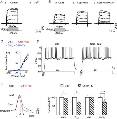
Aa and Ab, whole-cell recording of voltage-dependent currents from an amacrine cell in Ringer solution,100 μm Cd2+ + 1 μm TTX reduced both the K+ and Na+ currents. Ba, the addition of 100 μm picrotoxin and 10 μm strychnine, referred to as a Cd2+ inhibitory cocktail Cd(I), had little effect on the outward K+ currents. Bb, taurine markedly enhanced the K+ current amplitudes. Bc, the effect of taurine was insensitive to 10 μm CGP55845, a GABAB antagonist. C, the I–V curves measured and plotted from 8 cells in each of the test solutions. D, taurine increases the firing rate of APs generated by current injection (15 pA) in a ganglion cell superfused with the Cd(I) solution. Similar results were obtained from 7 of the 15 ganglion cells tested. The other 8 cells, in which injecting depolarizing currents generated spikes, rapidly accommodated and stopped firing within 1 s; these cells were not tested further. E, an expanded spike recording to indicate the points at which measurements were made of the peak, half-duration (D1/2), rise and decay times of APs in Cd(I) and with taurine (left). Bar graphs (right panel) show that taurine reduced the peak by 7.4 ± 2% (*P > 0.02, n = 189), D1/2 by 26.2 ± 7.2% (**P > 0.01, n = 189) and decay time by 27.1 ± 8.3% (***P > 0.01, n = 189).