
Figure 4. Taurine activates 5-HT2A receptors in retinal neurons.
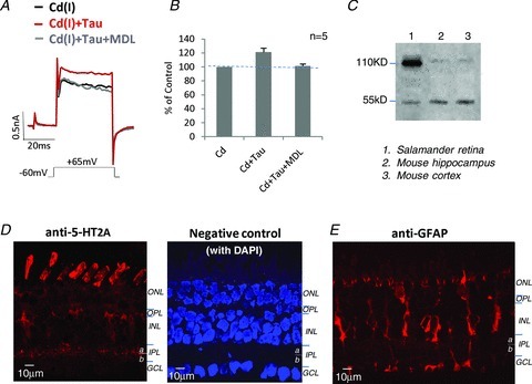
A, taurine enhanced KV currents in the Cd(I) solution, but the effect was blocked by 1 nm MDL11939, a selective 5-HT2A receptor antagonist. B, the bar graphs show the summary results of the effect of MDL11939 on blocked taurine-enhanced KV current amplitudes measured at +65 mV. C, Western blots show that the anti-5-HT2A detected protein bands at the predicted MW of 53 kDa in samples from mouse brain and salamander retina. It is likely that protein bands near MW of 110 kDa are dimers. D, confocal image of anti-5-HT2A labeling in a salamander retinal section, depicting the strong labeling in photoreceptors and within the inner plexiform layer (IPL); punctate labeling is also seen in the somas of the inner nuclear layer (INL) and ganglion cell layer (GCL); and the negative control (omitting 5-HT2A antibody) shows no labeling in retinal sections (the cell nuclei were stained with 4′,6-diamidino-2-phenylindole (DAPI)). E, Muller cells in a retinal section were labeled by the anti-glial fibrillary acidic protein (GFAP). ONL. outer nuclear layer; OPL, outer plexiform layer.