
Figure 5. Enhancement of LTP in the adult glutamate NMDAR subunit 3A (GluN3A) knockout (KO) brain.
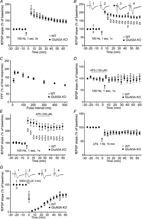
LTP induction in hippocampal slices from adult wild-type (WT) and GluN3A KO mice. A, LTP was induced by HFS (100 Hz, 1 s) repeated three times (3×). The magnitude of LTP was similar between WT and GluN3A KO slices. Each group: n = 6; B, LTP was induced by a single episode of HFS (100 Hz, 1 s, 1×). Although the initial elevated level of fEPSPs was similar, the magnitude of LTP in GluN3A KO slices stayed significantly higher than that in WT slices up to 60 min. WT and KO groups: n = 15 and 17, respectively; P < 0.05 between the averaged slopes of fEPSPs of WT and KO slices. C, paired-pulse facilitation (PPF) of fEPSP induced by various inter-pulse intervals was similar between adult WT and GluN3A KO slices. Both groups: n = 4. D, LTP was recorded in WT and GluN3A KO slices from adult animals in the presence of the NMDAR antagonist d(−)2-amino-5-phosphonovalerate (AP-5; 100 μm). LTP was similarly inhibited in both WT and GluN3A KO slices when AP-5 was bath applied 20 min prior to HFS (100 Hz, 1 s). Each group: n = 5. E, AP-5 (100 μm) application after the HFS (100 Hz, 1 s) stimulation could not block LTP induction in both WT and GluN3A KO slices. Each group: n = 5. F, similar LTD induced by LFS (1 Hz for 15 min) was recorded in WT and GluN3A KO slices from adult mice. Each group: n = 6. G, similar LTD was induced by 4 min exposure to 30 μm NMDA in WT and GluN3A KO slices from adult mice. Each group: n = 6.