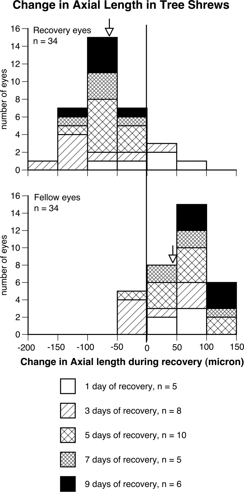
Figure 3.
The frequency distributions of change in axial length (from the anterior surface of cornea to the inner surface of sclera) in the treated eyes that recovered from deprivation-induced myopia for various durations (top) and in their untreated, fellow eyes within the same duration (below) in tree shrews. A total of 30 treated eyes shortened during recovery, whereas 5 fellow eyes shortened within the same duration (P < 0.0001, χ2 test).

