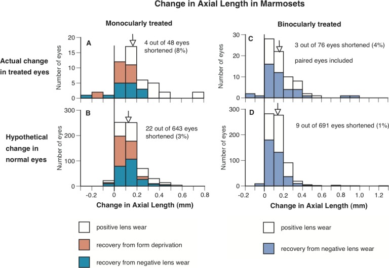
Figure 5.

The frequency distributions of change in axial length (from the anterior surface of cornea to the posterior surface of sclera) in the treated eyes of marmosets that either wore positive lenses, or recovered from form deprivation or wearing negative lenses (A, C), and in untreated eyes in normal animals grouped to match the same duration of visual exposure as the treated eyes through interpolation (B, D). Left and right represent the change in axial length in the eyes of monocularly- and binocularly-treated marmosets (and the corresponding interpolated untreated eyes), respectively. It is clear that a higher percentage of the treated eyes shortened (axial change below zero) than the calculated data from age-matched normal eyes. The vertical lines indicate where zero is on the x-axes, and the arrows indicate the mean change in axial length for each group of eyes.
