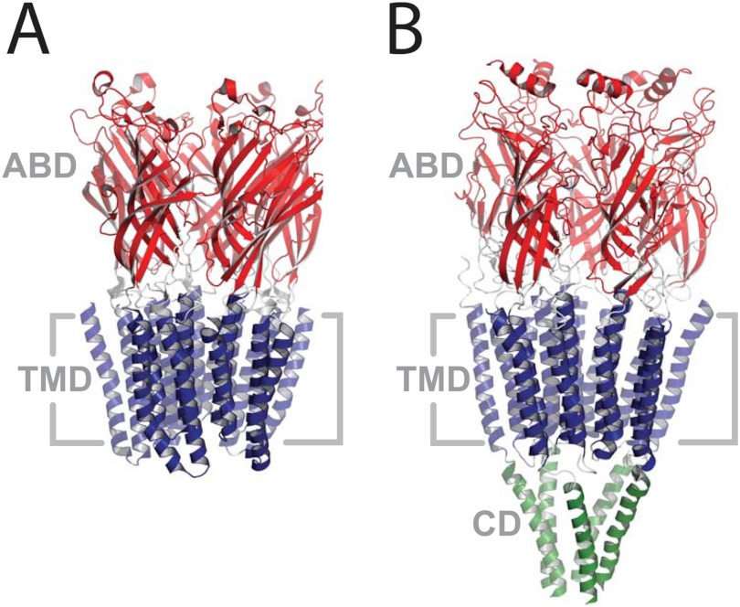
FIGURE 1.
The structures of GLIC (A, Protein Data Bank code 3EAM) and the Torpedo nAChR (B, Protein Data Bank code 2BG9). Both structures are side views from within the plane of the membrane. Coloring highlights the domain structure: the extracellular agonist-binding domain (ABD) in red, the transmembrane domain (TMD) in blue, and the cytoplasmic domain (CD) in green.

