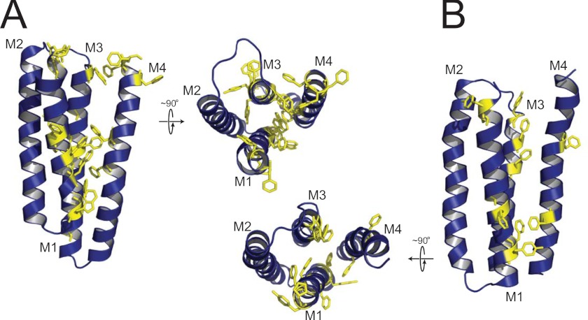
FIGURE 6.
Aromatic-aromatic interactions may dictate the propensity of a pLGIC to adopt a lipid-dependent uncoupled conformation. Shown is a comparison of the aromatic residues located in M1, M3, and M4 of a single subunit transmembrane domain for GLIC (A) and the α-subunit of the nAChR (B). Both a side view of the transmembrane domain of each subunit and a top view looking down at the bilayer surface are shown. Note that the side view orientations of the two transmembrane domains have been rotated 180° about the long axis of each molecule relative to the side view orientation shown in the schematic diagram of uncoupling in supplemental Fig. S6, because this presents a clearer view of the aromatic-aromatic interactions at the interface between M4 and M1+M3.

