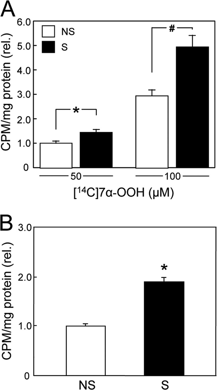
FIGURE 2.

Uptake of radioactive 7α-OOH by whole MA-10 cells and by their mitochondria. A, cells stimulated (S) with 1 mm Bt2cAMP for 3 h (cf. Fig. 1), along with nonstimulated (NS) controls, were incubated with 50 or 100 μm [14C]7α-OOH in POPC/Ch/[14C]7α-OOH (1.0:0.8:0.2 by mol) SUVs for 5 h and then washed thoroughly, recovered, and analyzed by scintillation counting. rel., relative. B, cells from the same stimulated (S) and nonstimulated (NS) populations in A were recovered after a 2-h exposure to 50 μm SUV-[14C]7α-OOH. Cells were homogenized, and Mito were isolated by differential centrifugation and analyzed by scintillation counting. A and B: means ± S.E. of values from three separate experiments are plotted. A, 50 μm: *, p < 0.05 (stimulated versus nonstimulated); 100 μm: #, p < 0.01 (stimulated versus nonstimulated). B, *, p < 0.01 (stimulated versus nonstimulated).
