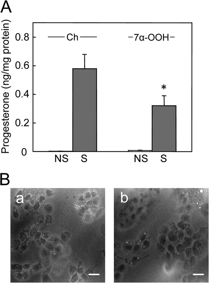
FIGURE 5.

Progesterone formation in 7α-OOH-challenged MA-10 cells. A, cells in a 96-well plate were preincubated overnight in serum-free DMEM/F12 medium. They were then either not treated (NS) or treated (S) with 1 mm Bt2cAMP in the presence of POPC/Ch/7α-OOH (0.8:1.0:0.2 by mol) SUVs (7α-OOH) or POPC/Ch (1:1 by mol) SUVs (Ch); total SUV lipid was 0.8 mm. After 3 h of incubation, the medium was removed and analyzed for progesterone by enzyme immunoassay, whereas cells were scraped into cold lysis buffer and analyzed for total protein. Plotted values are means ± S.E. (n = 3); *, p < 0.01 as compared with stimulated, Ch-treated. B, bright field microscopic images from experiment described in A. Stimulated cells were examined after being exposed to POPC/Ch (1:1 by mol) (panel a) or POPC/Ch/7α-OOH (0.8/1.0/0.2 by mol) (panel b) SUVs for 3 h. Bar: 75 μm.
