FIGURE 6.

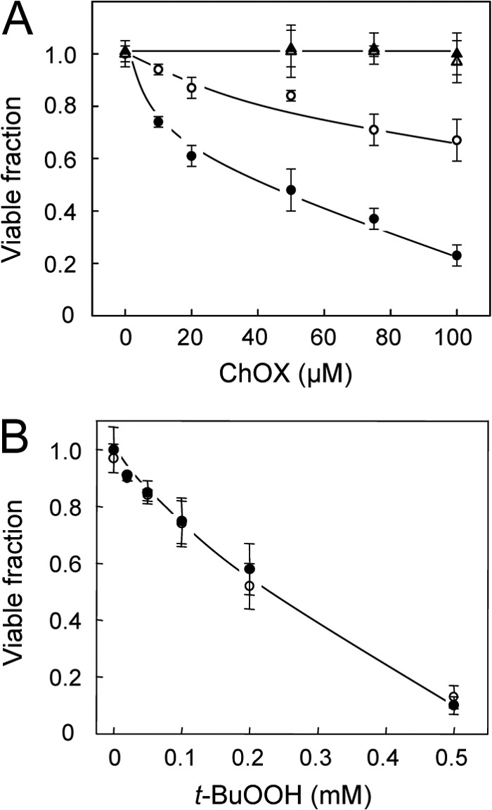
Effect of steroidogenic activation on MA-10 cell sensitivity to 7α-OOH versus t-BuOOH toxicity. A, cells at ∼70% confluency were incubated in the absence (nonstimulated) or presence (stimulated) of 1 mm Bt2cAMP for 1.5 h in serum-free medium, after which POPC/Ch/7α-OOH (1.0:0.5;0.5 by mol) or POPC/Ch/7α-OH (1.0:0.5:0.5 by mol) SUVs were introduced at the indicated cholesterol oxide (ChOX:7α-OOH or 7α-OH) concentration in bulk phase. B, similarly prepared cells were also challenged with t-BuOOH. After 16 h of incubation, cell viability was assessed by thiazolyl blue (MTT) assay. Data points are as follows: A, 7α-OOH, nonstimulated (○); 7α-OOH, stimulated (●); 7α-OH, nonstimulated (▵); 7α-OH, stimulated (▴). B, nonstimulated (○); stimulated (●). Means ± S.E. of values from 3–4 separate experiments are plotted in A and B.
