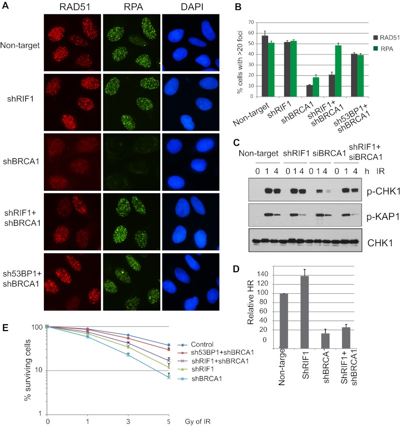
FIGURE 4.
Depletion of RIF1 alleviates DNA repair defect in BRCA1-knockdown cells. A, U2OS cells were infected with lentiviral particles carrying the indicated shRNAs. 48 h post-infection, cells were irradiated with 10 Gy IR and recovered for 4 h. Immunostaining was performed using antibodies as indicated. B, quantification of RAD51 and RPA foci formation in cells was as described in A. More than 150 cells were counted to determine the percentages of foci forming cells in each sample. C, checkpoint activation in single- or double-knockdown cells is shown. Control or RIF1 stable knockdown cells were transfected with control or BRCA1-specific siRNAs. 48 h later cells were exposed to 0 or 10 Gy IR and harvested at the indicated time points. Total lysates were subjected to Western blot analysis with the indicated antibodies. D, DR-GFP reporter (U2OS-DR) cells were infected with indicated lentivival shRNAs for 48 h and then electroporated with I-SceI expression plasmid (pCBASce). The percentage of GFP-positive cells was determined by flow cytometry 48 h after electroporation. The data were normalized to those obtained from cells infected with non-targeting shRNA. Data represent the means (±S.D.) of three independent experiments. E, shown is clonogenic survival of cells as described in A after exposed to the indicated doses of IR. Results are the means (±S.D.) of three independent experiments.

