
FIGURE 5.
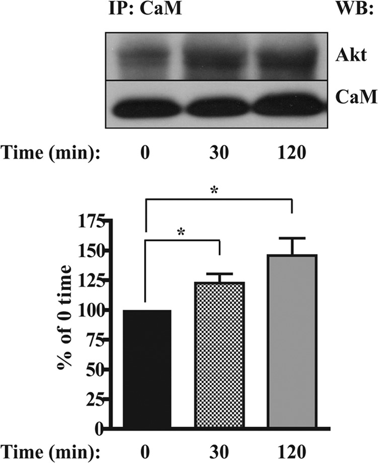
PDGF-BB-dependent co-immunoprecipitation of Akt with CaM. ST88-14 cells (1.5 × 10⁶/time point) were serum-starved for 4 h and then treated with 20 ng/ml PDGF-BB for 0 (untreated), 30, and 120 min. Cells were solubilized in lysis buffer, and the lysates were incubated overnight with anti-CaM antibody and precipitated with protein A/G-agarose. Immunoprecipitated Akt was detected by Western blotting (WB) using an antibody to total Akt. Results are mean ± S.E. from five independent experiments. *, p < 0.05. IP, immunoprecipitation.