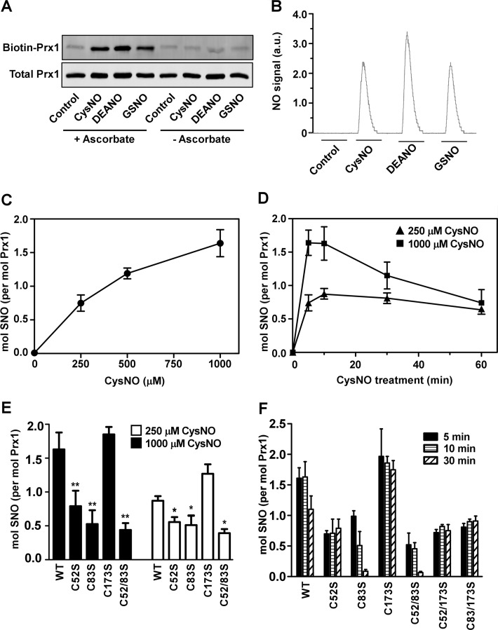
FIGURE 1.
S-Nitrosylation of Prx1 in vitro. A, fully reduced Prx1 was incubated for 10 min at 37 °C with CysNO (250 μm), DEANO (500 μm), or GSNO (250 μm). SNO-Prx1 was assessed by the biotin switch assay in the presence or absence of ascorbate. B, after treatment with different NO/SNO donors as in A, nitrosylation of Prx1 was assessed by chemical reductive chemiluminescence. a.u., arbitrary units. C, after incubation of fully reduced Prx1 with different concentrations of CysNO for 5 min at 37 °C, SNO content was measured by the Saville-Griess assay. D, Prx1 was incubated with 250 or 1000 μm CysNO for different times at 37 °C. SNO content was determined as in C. E, reduced Prx1, WT, or Cys to Ser mutants were incubated with 250 or 1000 μm CysNO for 10 min. SNO content was determined as in C. F, reduced Prx1, WT, or Cys to Ser mutants were incubated with CysNO (1000 μm) for the indicated times. SNO content was determined as in C. Results shown represent the mean ± S.D. n ≥ 3. *, p < 0.05; **, p < 0.01 for mutant versus WT by one-way analysis of variance with post hoc Tukey test.

