
FIGURE 1.
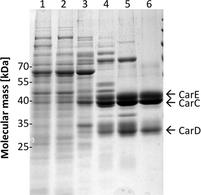
Purification of the caffeyl-CoA reductase-Etf complex. Samples from the different purification steps were separated by SDS-PAGE, and proteins were stained with Coomassie Brilliant Blue. Lane 1, cell extract; lane 2, cytoplasm; lane 3, pooled fractions from Q Sepharose; lane 4, pooled fractions from phenyl-Sepharose; lane 5, pooled fractions from Blue Sepharose; lane 6, pooled fractions from Sephacryl S-300. 10 μg of protein was applied to each lane.