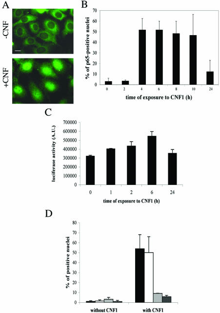
Figure 6.
Rac-induced activation of NF-κB. Fluorescence micrographs of HEp-2 cells untreated (-CNF1) or treated for 4 h with 10-9 M CNF1 (+CNF1), stained with an anti-p65 antibody. The illustrations are used to note the positively stained nuclei after CNF1 exposure. (B) Graph showing the time-dependent nuclear translocation of p65 after the CNF1-induced Rac activation. (C) Graph showing the luciferase activity induced by the CNF1-induced Rac activation on HEp-2 cells transfected with a plasmid corresponding to a luciferase reporter gene under the control of six repeated κB-binding sites. It is evident that NF-κB translocation correlated with its gene transactivation activity. (D) Graph showing the effects of the Rho family inhibiting toxins on the CNF1-induced p65 nuclear translocation. Cells were preexposed to toxins for 2 h before addition of CNF1 for an additional 4 h. Note that the two Rac-inhibiting toxins (LT82, which inhibits Rac, and LT9048, which inhibits Rac and Cdc42) dramatically reduce the percentage of p65-positive nuclei, whereas C3B, the Rho-inhibiting toxin, does not. (▪, control cells; □, C3B-treated cells;  , LT82-treated cells; and
  , LT9048-treated cells).

