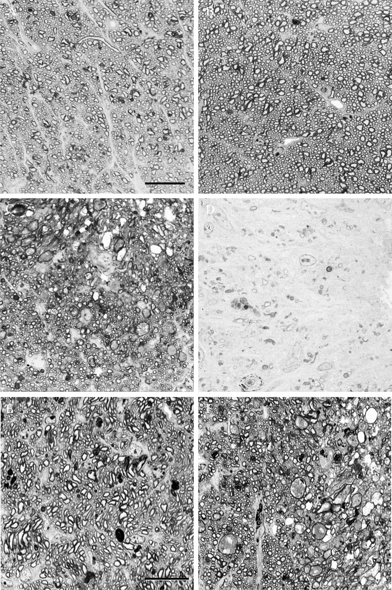
Figure 3. .
High-magnification photomicrographs showing optic nerve sections from the control eye (A) and optic nerve crush eyes that were graded as having mild (B), moderate (C), and severe (D) optic nerve damage. Scale bar: 20 μm. Moderate optic nerve damage in experimental glaucoma (E) and optic nerve crush (F). Scale bar: 20 μm.

