Figure 8.
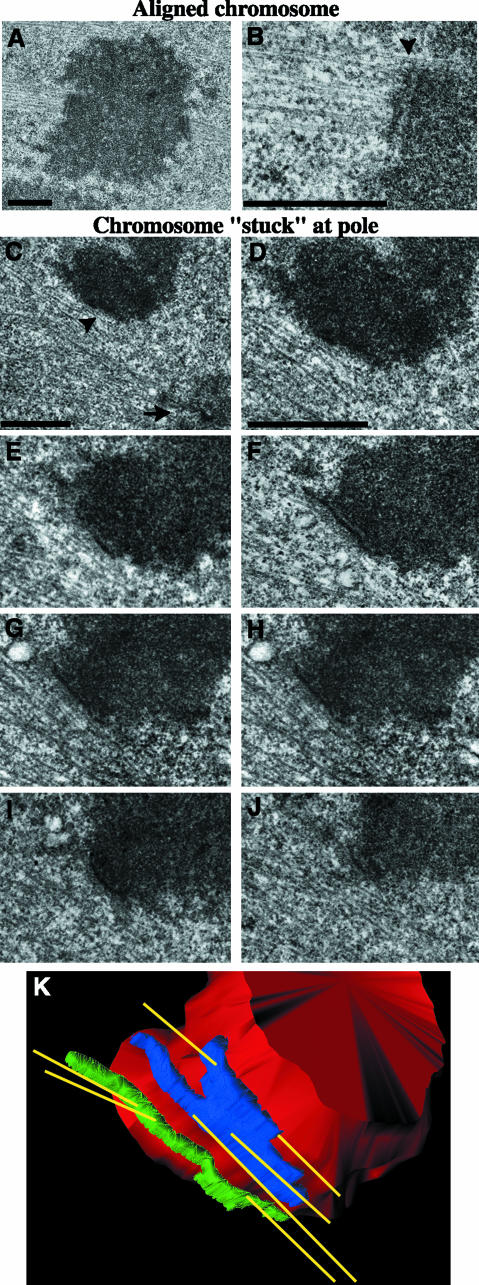
Centromeric MCAK depletion leads to improper kinetochore–microtubule attachments in PtK2 cells. Correlative light/serial-section EM was performed on GFP-CEN–injected cells to analyze kinetochore and K-fiber morphology. Low-magnification (A) and high-magnification (B) images of an aligned chromosome with normal kinetochore morphology and MT attachments; arrowhead is positioned at the kinetochore–MT interface. (C) Low-magnification image of a chromosome that remained for an extended period of time at the pole during mitosis. Arrow denotes a centrosome; arrowhead denotes a kinetochore. (D–J) High-magnification, 100-nm serial-section images of the chromosome shown in C reveal that both kinetochores are attached to MTs emanating from both poles, which makes this chromosome both merotelically and syntelically oriented on the spindle. (K) The 3-D structure of MTs attached to the merotelic-syntelic kinetochores of the chromosome shown in C–J. The chromosome is shown in red, MTs in yellow, and sister kinetochores in blue and green. Bars, 1 μm.

